Centeno CJ; Schultz J; Freeman M. Sclerotherapy of Baker’s cyst with imaging confirmation of resolution. Pain Physician (United States), Mar-Apr 2008, 11(2) p257-61
Bakers Cyst: Dextrose + Sodium Morrhuate: Centeno 2008
Dr. Reeves’ Notes: This study demonstrated a case of MRI use to demonstrate shrinking of a Baker’s cyst by simply injecting the knee, since the knee lining is connected with the Baker’s cyst. This suggests a favorable change in the joint to stop overproduction of joint fluid which allows the cyst to shrink since it is connected with the knee joint. Simple intra articular injection of 12.5% dextrose and sodium morrhuate on 3 occasions in a patient who failed conservative care and drainage was described. In the present and near future both MRI and ultrasound scans can be used to objectively document in this way. Anticipate more such studies.
A PDF version of the Bakers Cyst study is available here.
An abstract of the study on Bakers Cyst is available here, with a copy of the content below…
BACKGROUND:
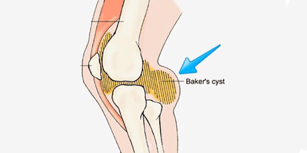
Baker’s cysts are commonly encountered in pain management practices.
OBJECTIVE:
To ascertain if sclerotherapy treatment of a Baker’s cyst could produce objectively verifiable MRI imaging changes.
DESIGN:
Case report.
METHODS:
A 52-year-old white male with a posterior horn of the medial meniscus tear and a large Baker’s cyst who had failed conservative care and drainage was imaged before treatment with sclerosing. Three injections of 12.5% dextrose and anesthetic with sodium morrhuate were injected intraarticular into the right knee after drainage.
RESULTS:
The Baker’s cyst resolved on both postoperative imaging after the completion of care as well as on physical examination.
CONCLUSIONS:
Prolotherapy in this case study seemed to be an effective treatment for Baker’s cyst in this patient.
AUTHOR’S ADDRESS:
The Centeno-Schultz Clinic Westminster, CO, USA. (centenooffice@centenoclinic.com)


